Table of Contents    
Original Article
 
Ultrastructure of ovarian preantral follicles and corpus luteum in Indian flying fox Pteropus giganteus (Brünnich)
Ajay V Dorlikar1, Amir A Dhamani2, Pravin N Charde3, Anil S Mohite3
1MSc (Zoology) Research Scholar, PG Department of Zoology and Research Academy, Sevadal Mahila Mahavidyalaya, Sakkardara Square, Nagpur, Maharashtra, India.
2MSc (Zoology) PhD, PG Department of Zoology and Research Academy, NH College, Bramhapuri, Maharashtra, India.
3MSc (Zoology) PhD, PG Department of Zoology and Research Academy, Sevadal Mahila Mahavidyalaya, Sakkardara Square, Nagpur, Maharashtra, India.

Article ID: 100001C06AD2014
doi:10.5348/c06-2014-1-OA-1

Address correspondence to:
Ajay Vasantrao Dorlikar
PG, Department of Zoology and Research Academy
Sevadal Mahila Mahavidyalaya, Sakkardara Square
Nagpur, Maharashtra
India
Phone: 091-9422809685
Fax: 091-0712-2705037
Email: ajaydorlikar@gmail.com

Access full text article on other devices

  Access PDF of article on other devices

[HTML Abstract]   [PDF Full Text] [Print This Article]
[Similar article in Pumed] [Similar article in Google Scholar]

How to cite this article
Dorlikar AV, Dhamani AA, Charde PN, Mohite AS. Ultrastructure of ovarian preantral follicles and corpus luteum in Indian flying fox Pteropus giganteus (Brünnich). Edorium J Cell Biol 2014;1:1–11.


Abstract
Aims: This study was undertaken to reveal the ultrastructural details of preantral ovarian follicles and corpus luteum in Indian flying fox Pteropus giganteus.
Methods: Five specimens were collected during estrous, early pregnancy and mid-pregnancy from the feeding sites. Reproductive stages were confirmed by the histological examination of the uterus and ovaries. The ultrastructural details in the preantral follicle and luteal cells were studied using transmission electron microscope.
Results: Primordial graffian follicle (type-1) was composed of an oocyte surrounded by incomplete layer of 4–5 flattened granulosa cells resting on the basement membrane. In primary follicles oocyte was encircled by single layer of 10–12 cuboidal granulosa cells. Oocyte possesses numerous bent microvilli. In small preantral follicle (type-3), complete two to four layers of cuboidal granulosa cells were noted. Zona pellucida appears at this stage. Large preantral follicles (type-4) possessed four to six layers of granulosa cells resting on basement membrane. Single extrovert type of corpus luteum was observed during early to mid-pregnancy. Diameter of large luteal cells during early and mid pregnancy were 22.46±0.56 µm and 22.85±0.56 µm and of small luteal cells were 17.81 ± 0.40 µm and 17.34 ± 0.46 µm respectively.
Conclusion: Biphasic pattern of development of preantral follicle had been observed in Pteropus giganteus. Oocyte of large preantral follicles (type-4) was characterized by the presence of cortical granules in the cortical region. Ultrastructural features of large and small luteal cells confirms to the criteria of steroid secreting cells. No significant differences were noted in the diameter of large and small luteal cell during early and mid-pregnancy.

Keywords: Corpus luteum, Luteal cell, Oocyte, Pteropus giganteus, Preantral follicle


Introduction

Bats comprise more than 20% of the mammalian species of the world. [1] The study of the bat reproduction is of immense importance due to variety of adaptations possessed by them in response to environmental conditions in which they live. Bats living in temperate regions, show heterothermy and hibernate during winter for successful survival. For successful reproduction bats show delayed ovulation, delayed implantation and sperm storage like phenomenon. [2] [3] Special mechanism of flight in bats has improved the survival of bats by improving the ability to exploit variety of natural resources. [4] [5] [6] Thus, the investigations on the function of the ovary is of great importance to elucidate the characteristics of reproductive pattern of bats. Ovary is the principle sex organ responsible for oogenesis and secretion of steroid hormones. During oogenesis, maturation and development of graffian follicle as well as ultrastructure of granulosa cells is a matter of interest due to role of granulosa and theca cells in the secretion of oestradiol. After luteinization the luteal cells are primarily responsible for production of progesterone which is responsible for maintaining pregnancy. Information regarding ultrastructure of preantral follicles and luteal cells in Pteropus giganteus is very scarce. To understand the reproductive physiology of Pteropus giganteus, we attempted to study the ultrastructural details in the preantral follicle and luteal cells.


Materials and Methods

Collection of specimens
All the specimens used during entire study period were obtained from natural populations from Padmapur village, Longitude 20°22'N and latitude 79°48'E, (Dist-Chandrapur, Maharashtra) India. The five specimens were collected during estrous, early pregnancy and mid-pregnancy from the feeding sites. Reproductive stages were confirmed by the histological examination of uterus and ovaries. Live specimens were brought to the laboratory with minimum stress and then killed by decapitation. The work was approved by the ethics committee of institute.

Procedure for transmission electron microscopy
The ovaries were dissected out, freed from extraneous fat and fixed in 2.5% glutaraldehyde and paraformaldehyde prepared in cacodylate buffer (0.1 M, pH 7.4) adjusted to pH 7.2. The ovaries were kept in the fixative solution for 24 hours at 4°C. After 24 hours, the tissues were rinsed in cacodylate buffer and then post-fixation was done for three hours in 1% 0.067 M cacodylate-buffered osmium tetroxide. Tissues were treated with a varying concentration of ethyl alcohol for dehydration. Dehydrated tissues were cleared in propylene oxide solution. Then the sections were blocked in embedding moulds with Araldite CY-212, dodecenyl succinic anhydride (DDSA) and benzyldimethylamine (BDMA) which polymerize at 60°C. Semi-thin sections were seen under the microscope after staining with toluidine blue. Then, ultrathin sections from selected blocks were cut with glass knife (300–700 Å thick) and were double stained with 10% alcoholic uranyl acetate for 20 minutes and in Reynold's lead citrate for 10 minutes. Sections were picked up on 400-mesh copper grids. The sections were examined and electron photo micrographs were taken by transmission electron microscope (JEOL-100 S) at 80 KV accelerating voltage.

Statistical analysis
A statistical analysis of data was performed to determine the significant changes in large and small luteal cells during early and mid-pregnancy. Mean, standard error, standard deviation, variance and ANOVA with post hoc Tucky's HSD test were calculated by using Statistical Package for Social Sciences (SPSS 10.0).


Results

Ultrastructure of ovarian preantral follicles
The study revealed that primordial graffian follicle (type-1) was composed of an oocyte surrounded by incomplete layer of 4–5 flattened and poorly differentiated granulosa cells resting on the basement membrane (Figure 1). Oocyte of primordial follicles had round nuclei. Nucleus was located in central or eccentric position and possessed reticulated nucleoli. Oocyte had large pleomorphic mitochondria and well-developed golgi complexes. Vesicles were also observed in close association with mitochondria. Mitochondria observed in the ooplasm were round with longitudinal cristae while some mitochondria were elongated with transverse cristae. Lipid droplets, smooth endoplasmic reticulum and rough endoplasmic reticulum were present in all oocytes. Microvilli were present on the oolemma. Gap junctions between granulosa cells and microvilli were visible in between oolemma and granulosa cells (Figure 2) (Figure 3). In the successive development primordial follicle grows and becomes primary follicle (type-2). In our study this stage was characterized by change in histology of granulosa cells. In primary follicles oocyte was encircled by single layer of 10–12 cuboidal granulosa cells. Oocyte possessed numerous bent microvilli. Rounded mitochondria were present. Lipid droplets were also seen. Smooth endoplasmic reticulum and rough endoplasmic reticulum along with free ribosomes were also seen (Figure 4) (Figure 5) (Figure 6).

In small preantral follicles (type-3) complete two to four layers of cuboidal granulosa cells were noted. Cell organelles of the oocyte of these follicles were observed towards the cortical region, away from the nucleus. Zona pellucida appears at this stage. These follicles had a rich blood supply (Figure 7). Large preantral follicles (type-4) possessed four to six layers of granulosa cells resting on the basement membrane. Theca layer was observed in the vicinity of the basement membrane (Figure 8). Ooplasm possessed agranular endoplasmic reticulum and mitochondria with transverse cristae (Figure 9). Large preantral follicles were associated with other preantral follicles (Figure 10). In healthy follicle, granulosa layer is separated from lamina densa by lamina lucida. Continuous lamina densa indicates the healthy status of the follicle. The reticular layer that separates theca from lamina densa is lamina lucida filled with fibrils which are made up of collagen (Figure 11). Theca interna cell possessed rounded mitochondria and chromatin material was observed at the peripheral region of the nucleus (Figure 12). Oocyte showed all the cell organelles along with cortical granules in the cortical region.

Ultrastructure of corpus luteum
In Pteropus giganteus, single extrovert type of corpus luteum had been observed. During early to mid-pregnancy significant increase in size of corpus luteum has been observed. At mid-pregnancy corpus luteum occupies two-third portion of the ovary. During early and mid-pregnancy corpus luteum appears active and secretory. Corpus luteum possessed discrete population of large and small luteal cells with blood vessels. Large luteal cells had centrally placed nuclei and distinct nucleoli (Figure 13) (Figure 14). Diameter of large luteal cell during early and mid-pregnancy was found to be 22.46±0.56 µm and 22.85±0.56 µm, respectively. In the vicinity of large luteal cells, small luteal cells were noted having prominent nucleus and nucleoli. Diameter of small luteal cell during early and mid-pregnancy was found to be 17.81±0.40 µm and 17.34±0.46 µm, respectively. No significant differences were noted in the diameter of large and small luteal cell during early and mid-pregnancy (Table 1).

Large luteal cells
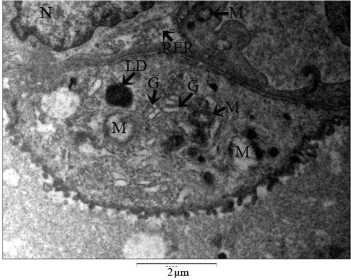
The plasma membrane of large luteal cells had cytoplasmic projections which connects it to either large or small luteal cell. Plasmalemma showed the pinocytic vesicles through which transportation of secretions occurs in the cell (Figure 13) (Figure 14) (Figure 15). Rounded or elongated mitochondria with tubular cristae were dispersed in the cytoplasm of the cell. Smooth endoplasmic reticulum and rough endoplasmic reticulum were present in the cytoplasmic matrix. Smooth endoplasmic reticulum was connected together with interconnecting tubules. Polyribosomes were observed in the cytoplasm of these cells (Figure 16). Golgi complexes were also present in the cytoplasm of large luteal cells. Golgi apparatus was found to be in association of vesicles. Discontinuous cisternae were noted in the Golgi complex. Electron dense, opaque, lipid droplets were prominently present in the cytoplasm. Few electron dense to electron lucent secretory vesicles were observed in the cytoplasmic matrix (Figure 17) (Figure 18) (Figure 19).

Small luteal cells
Ultrastructure of small luteal cells resemble that of large luteal cells (Figure 20) (Figure 21). In the cytoplasmic matrix of these cells, mitochondria were elongated to oval in shape with transverse cristae. Vesicles along with Golgi complex were reported in the cytoplasm. Plasma membrane of these cells also showed cytoplasmic projections for making connections with the neighbouring cells. Smooth and rough endoplasmic reticulum with few cisternae was also present in the cytoplasm. Electron dense opaque lipid droplets were also observed in these cells (Figure 22) (Figure 23) (Figure 24).

Cursor on image to zoom/Click text to open image
Figure 1: Primordial follicle in the ovary of Pteropus giganteus showing oocyte encircled by incomplete layer of flattened follicular cells on the basement membrane. (Glutaraldehyde/osmium tetroxide, x2,500).
BM: Basement membrane, GC: Granulosa cell.


Cursor on image to zoom/Click text to open image
Figure 2: Primordial follicle showing presence of microtubules in the cytoplasm of the oocyte. Gap junctions between granulosa cells and microvilli are visible in between oolemma and granulosa cells. (Glutaraldehyde/osmium tetroxide, x5,000).
BM: Basement membrane, GC: Granulosa cell, GP: Gap Junction, M: Mitochondria, Mv: Microvilli, O: Oolemma, V: Vacuoles.


Cursor on image to zoom/Click text to open image
Figure 3: Cytoplasmic organelles in the oocyte of primordial follicle. (Glutaraldehyde/ osmium tetroxide, x10,000).
Mt: Microtubules, M: Mitochondria, Mv: Microvilli, V: Vacuoles.


Cursor on image to zoom/Click text to open image
Figure 4: Arrangement and shape of the granulosa cells. Granulosa cells resting on the basement membrane. (Glutaraldehyde/osmium tetroxide, x3,000).
BM: Basement membrane, GC: Granulosa cell, N: Nucleus.


Cursor on image to zoom/Click text to open image
Figure 5: Cytoplasmic organelles in the oocyte of Primordial Follicle. (Glutaraldehyde/osmium tetroxide, x40,000).
M: Mitochondria, RER: Rough endoplasmic reticulum, Mv: Microvilli.


Cursor on image to zoom/Click text to open image
Figure 6: Detail of peripheral cell of the granulosa. (Glutaraldehyde/osmium tetroxide, x10,000).
N: Nucleus, M: Mitochondria, O: Oocyte, LD: Lipid droplet.


Cursor on image to zoom/Click text to open image
Figure 7: Arrangement and shape of the granulosa cells in small preantral follicle. Note the Granulosa cells in the vicinity of blood vessel. (Glutaraldehyde/osmium tetroxide, x2,000).
BM: Basement membrane, BV: Blood vessel, GC: Granulosa cell, N: Nucleus.


Cursor on image to zoom/Click text to open image
Figure 8: Arrangement and shape of the granulosa cells in large preantral follicle. Granulosa cells resting on the basement membrane. (Glutaraldehyde/osmium tetroxide, x3,000).
BM: Basement membrane, BV: Blood vessel, GC: Granulosa cell, N: Nucleus, TI: Theca interna.


Cursor on image to zoom/Click text to open image
Figure 9: Cytoplasmic organelles in the granulosa cell of large preantral follicle. (Glutaraldehyde/osmium tetroxide, x40,000).
CG: Cortical granule, M: Mitochondria, N: Nucleus, RER: Rough endoplasmic reticulum.


Cursor on image to zoom/Click text to open image
Figure 10: Arrangement and association of the preantral follicles. Granulosa cells resting on the basement membrane. Note theca interna outside the basement membrane (Glutaraldehyde/osmium tetroxide, x2,500).
BM: Basement membrane, BV: Blood vessel, GC: Granulosa cell, N: Nucleus, TI: Theca interna.


Cursor on image to zoom/Click text to open image
Figure 11: STheca interna outside the basement membrane of the preantral follicles. (Glutaraldehyde/osmium tetroxide, x4,000). BM: Basement membrane, TI: Theca interna.


Cursor on image to zoom/Click text to open image
Figure 12: Details of theca interna cell of the preantral follicles. (Glutaraldehyde/osmium tetroxide, x10,000).
M: Mitochondria, N: Nucleus, TI: Theca interna.


Cursor on image to zoom/Click text to open image
Table 1: Comparison of diameter of small and large luteal cells during early and mid- pregnancy in Pteropus giganteus. HSD [0.05] = 1.92; HSD [0.01] = 2.39.


Cursor on image to zoom/Click text to open image
Figure 13: Arrangement of large and small luteal cells during early pregnancy. (Glutaraldehyde/osmium tetroxide, x2,000). LLC: Large luteal cell, N: Nucleus, SLC: Small luteal cell.


Cursor on image to zoom/Click text to open image
Figure 14: Arrangement of large and small luteal cells during early pregnancy (Glutaraldehyde/osmium tetroxide, x2,500). Note indented nucleus of small luteal cell.
LLC: Large luteal cell, N: Nucleus, SLC: Small luteal cell.


Cursor on image to zoom/Click text to open image
Figure 15: Plasma membrane of large luteal cells during early pregnancy showing cytoplasmic projections. (Glutaraldehyde/osmium tetroxide, x6,300).
CP: Cytoplasmic projections, N: Nucleus.


Cursor on image to zoom/Click text to open image
Figure 16: Details of cytoplasmic organelles of large luteal cells during early pregnancy. Note the blood vessel in the vicinity of luteal cell, hypertrophied mitochondria with vesicular cristae and many lipid droplets (Glutaraldehyde/osmium tetroxide, x5,000).
BV: Blood Vessel, LD: Lipid droplet, M: Mitochondria.


Cursor on image to zoom/Click text to open image
Figure 17: Details of cell organelles of large luteal cells during early pregnancy. Note the oval hypertrophied mitochondria with tubular cristae, Dark and light osmiophilic lipid droplets, short tubular cisternae of RER, (Glutaraldehyde/osmium tetroxide, x4000).
LD: Lipid droplet, M: Mitochondria, N: Nucleus, NP: Nuclear pore, RER: Rough endoplasmic reticulum.


Cursor on image to zoom/Click text to open image
Figure 18: Details of cell organelles of Large luteal cells during early pregnancy. Note numerous Golgi complexes, oval hypertrophied mitochondria with tubular cristae, Dark osmiophilic lipid droplets, short tubular cristae of RER (Glutaraldehyde/osmium tetroxide, x3200).
Abbreviation: G - Golgi bodies, LD - Lipid droplet, M - Mitochondria, N - Nucleus, RER - Rough endoplasmic reticulum.


Cursor on image to zoom/Click text to open image
Figure 19: Details of cell organelles of large luteal cells during mid pregnancy. Note oval to globular mitochondria with lamellar to vesicular cristae. Dark and light osmiophilic lipid droplets, Tubular cisternae of SER, Short tubular cisternae of RER and free ribosomes scattered in the cytoplasm. (Glutaraldehyde/osmium tetroxide, x8000).
M: Mitochondria, N: Nucleus, RER: Rough endoplasmic reticulum, SER: Smooth endoplasmic reticulum.


Cursor on image to zoom/Click text to open image
Figure 20: Ultrastructure of small luteal cells during early pregnancy. Note numerous dark osmiophilic lipid droplets, oval hypertrophied mitochondria and tubular cristae of RER (Glutaraldehyde/osmium tetroxide, x2,500).
LD: Lipid droplet, M: Mitochondria, N: Nucleus, RER: Rough endoplasmic reticulum.


Cursor on image to zoom/Click text to open image
Figure 21: Ultrastructure of small luteal cells during mid-pregnancy. Note numerous oval hypertrophied mitochondria with tubular cristae and lipid droplets. (Glutaraldehyde/osmium tetroxide, x1,600).
LD: Lipid droplet, M: Mitochondria, N: Nucleus, RER: Rough endoplasmic reticulum.


Cursor on image to zoom/Click text to open image
Figure 22: Details of cell organelles of small luteal cells during early pregnancy. Note numerous Golgi complexes, oval hypertrophied mitochondria with tubular cristae, dark osmiophilic lipid droplets, tubular cristae of RER and short cristae of SER (Glutaraldehyde/osmium tetroxide, x8,000).
G: Golgi bodies, LD: Lipid droplet, M: Mitochondria, RER: Rough endoplasmic reticulum, SER: Smooth endoplasmic reticulum.


Cursor on image to zoom/Click text to open image
Figure 23: Details of cell organelles of small luteal cells during mid pregnancy. Note oval mitochondria, Golgi complexes, dark osmiophilic lipid droplets, tubular cristae of RER and short cristae of SER. (Glutaraldehyde/osmium tetroxide, x4,000).
G: Golgi bodies, LD: Lipid droplet, M: Mitochondria, N: Nucleus, RER: Rough endoplasmic reticulum, SER: Smooth endoplasmic reticulum.


Cursor on image to zoom/Click text to open image
Figure 24: Details of cell organelles of small luteal cells during mid pregnancy. Note oval mitochondria, Golgi complexes, dark osmiophilic lipid droplets, tubular cristae of RER and short cristae of SER. (Glutaraldehyde/osmium tetroxide, x8000).
G: Golgi bodies, LD: Lipid droplet, M: Mitochondria, N: Nucleus, RER: Rough endoplasmic reticulum.


Discussion

Ultrastructure of oocytes and preantral follicles had been observed in many mammalian species. Hertig et al. [7] and Bruin et al. [8] in human, Zamboni et al. [9] in Rhesus monkey, Tassell et al. [10] and Matos et al. [11] in sheep, Fair et al. [12] in cow, Frankenberg and Selwood [13] in bushtail possum and Bielanska et al. [14] in pig had studied the ultrastructure of ovarian follicles. In Pteropus giganteus both the ovaries contain a variety of follicular types. Preantral follicles (type-1, type-2, type-3 and type-4) were observed in the ovary in ovarian cortex. In all follicular types the oocyte nucleus was either centrally or eccentrically located and most of the organelles were evenly distributed throughout the cytoplasm. Nuclear pores were observed in the nuclear membrane. Agranular endoplasmic reticulum was abundant. while granular endoplasmic reticulum was less so in the cytoplasm. Free ribosomes were present in the cytoplasm of all follicular types and Golgi complexes were observed. Oval to round mitochondria were noted. However, few mitochondria were elongated with transverse cristae. Vesicles were most abundant in the type-1 follicles. Lipid droplets were observed in most oocytes. Cortical granules were observed in the type-3 follicles and were located towards the oocyte periphery. Microvilli were present in all follicles, but appeared to increase in number and length as zona pellucida volume increased.

The corpus luteum is an endocrine gland which secretes large amounts of steroid hormones especially progesterone which prepares the uterine endometrium for implantation and maintains the early pregnancy. If fertilization and implantation do not occur, the ovulatory cycle ends and the corpus luteum undergoes luteolysis. The ultrastructure of corpus luteum of bat has been studied in Macrotus californicus [15] [16], Miniopterus schreibersii [17], Miniopterus schreibersii fuliginosus [18] and Taphozous longimanus [19]. Gopalkrishna et al. [20]had noted the extrovert corpus luteum in Megaderma lyra, Hipposideros fulvus and H. speoris. Anand Kumar [21] in Rhinopoma kinneri, Gopalkrishna et al. [22] in Hipposideros speoris, Sapkal et al. [23] and Seraphim et al. [24] in Hipposideros lankadiva had observed the extrovert type of corpus luteum. Corpus luteum shows two types of lutein cells called large luteal cells and small luteal cells.

The large luteal cells are derived from the granulosa cells while small luteal cells are derived from theca interna cells. [25] These cells were richly supplied by blood vessels. Large and small luteal cells actively participate in steroidogenic activity from early mid-pregnancy. In late pregnancy corpus luteum regresses. Morphological features of large luteal cells are comparable to those described for steroid secreting cells. [26] Large concentric whorls of granular endoplasmic reticulum were not observed. [27] Electron dense granules were reported in the cytoplasm. Few dense granules were observed in extracellular spaces with dense core and lighter periphery. A small luteal cell also conforms to the criteria of steroid secreting cells described by Christensen et al.[26] The features of a steroid-secreting cell described by these authors include abundant tubular agranular endoplasmic reticulum, lipid droplets and dispersed Golgi elements. These features were present in the cytoplasm of the small luteal cells of the Pteropus giganteus.

Cholesterol from low-density lipoprotein and high-density lipoprotein act as a precursor for synthesis of steroid hormones in graffian follicle. [28] Progesterone synthesis occurs by cholesterol side chain cleavage catalyzed by cytochrome P450scc in mitochondria to form pregnenolone which enters the endoplasmic reticulum and progesterone is synthesized in presence of 3-β-hydroxysteroid dehydrogenase. [29] Progesterone regulates its own synthesis by affecting the activity of the enzymes involved in steroidogenesis. [30] Thus increase in mitochondria and smooth endoplasmic reticulum in luteal cells during mid-pregnancy can be directly correlated with the increased level of progesterone in plasma during mid-pregnancy.


Conclusion

Biphasic pattern of development of preantral follicle was observed in Pteropus giganteus. Cell organelles present in large and small luteal cells during early and mid-pregnancy resemble that of steroid secreting cells. Thus both the cells might be playing significant role in the progesterone synthesis for maintenance of pregnancy. Abundant hypertrophied mitochondria, Golgi complexes and light osmophilic lipid droplets in the large and small luteal cells during mid-pregnancy indicate the increased steroidogenic process during mid-pregnancy.


References
  1. Purohit A, Vyas KB, Senacha KR. Population dynamics of bats in and around Jodhpur of great Indian desert. Tigerpaper 2006;33(3):15–22.    Back to citation no. 1
  2. Heideman PD. Delayed development in Fischer's pygmy fruit bat, Haplonycteris fischeri, in the Philippines. Journal of Reproduction and Fertility 1989;85(2):363–82.   [CrossRef]   [Pubmed]    Back to citation no. 2
  3. Sharifi M, Ghiorbani R, Fazeli A, Holt W. Evidance of sperm storage in Pipistrellus kuhlii (Chiroptera: Vespertilionidae) in western Iran. Folia zool 2004;53(1):1–6.    Back to citation no. 3
  4. Findley JS. Bats, a community perspective. Cambridge University Press 1993; P.167.    Back to citation no. 4
  5. Holmes DJ, Austad SN. Fly now, die later: Life-history correlates of gliding and flying in mammals. J Mammal 1994;75:224–6.   [CrossRef]    Back to citation no. 5
  6. Patterson BD, Willig MR, Stevens RD. Trophic strategies, niche partitioning, and patterns of Ecological Organization. In Bat Ecology. Edited by Kunz, T.H. & M.B. Fenton. The University of Chicago Press 2003; P. 536–79.    Back to citation no. 6
  7. Hertig AT, Adams EC. Studies on the human oocyte and its follicle. I. Ultrastructural and histochemical observations on the primordial follicle stage. J Cell Biol 1967;34(2):647–75.   [CrossRef]   [Pubmed]    Back to citation no. 7
  8. De Bruin JP, Dorland M, Spek ER, et al. Ultrastructure of the resting ovarian follicle pool in healthy young women. Biol Reprod 2002;66(4):1151–60.   [CrossRef]   [Pubmed]    Back to citation no. 8
  9. Zamboni L. Fine morphology of the follicle wall and follicle cell-oocyte association. Biol Reprod 1974;10(2):125–49.   [CrossRef]   [Pubmed]    Back to citation no. 9
  10. Tassell R, Kennedy JP. Early follicular development and atretic changes in the ovary of the lamb-- fine structure and histochemistry. Aust J Biol Sci 1980;33(6):675–87.   [Pubmed]    Back to citation no. 10
  11. Matos MH, Andrade ER, Lucci CM, et al. Morphological and ultrastructural analysis of sheep primordial follicles preserved in 0.9% saline solution and TCM 199. Theriogenology 2004;62(1-2):65–80.   [CrossRef]   [Pubmed]    Back to citation no. 11
  12. Fair T, Hulshof SC, Hyttel P, Greve T, Boland M. Oocyte ultrastructure in bovine primordial to early tertiary follicles. Anat Embryol (Berl) 1997;195(4):327–6.   [CrossRef]   [Pubmed]    Back to citation no. 12
  13. Frankenberg S, Selwood L. Ultrastructure of oogenesis in the brushtail possum. Mol Reprod Dev 2001;58(3):297–306.   [CrossRef]   [Pubmed]    Back to citation no. 13
  14. Bielanska-Osuchowska Z. Oogenesis in pig ovaries during the prenatal period: ultrastructure and morphometry. Reprod Biol 2006;6(2):161–93.   [Pubmed]    Back to citation no. 14
  15. Bleier WJ. Early embryology and implantation in the California leaf-nosed bat, Macrotus californicus. Anat Rec 1975;182(2):237–53.   [CrossRef]   [Pubmed]    Back to citation no. 15
  16. Crichton EG, Hoyer PB, Krutzsch PH. Cellular composition and steroidogenic capacity of the ovary of Macrotus californicus (Chiroptera: Phyllostomatidae) during and after delayed embryonic development. Cell Tissue Res 1990;260(2):355–66.   [CrossRef]   [Pubmed]    Back to citation no. 16
  17. Crichton EG, Seamark RF, Krutzsch PH. The status of the corpus luteum during pregnancy in Miniopterus schreibersii (Chiroptera: Vespertillionidae) with emphasis on its role in developmental delay. Cell Tissue Res 1989;258(1):183–201.   [CrossRef]   [Pubmed]    Back to citation no. 17
  18. Uchida TA, Inoue C, Kimura K. Effects of elevated temperatures on the embryonic development and corpus luteum activity in the Japanese long-fingered bat, Miniopterus schreibersii fuliginosus. Journal of Reproduction and Fertility 1984;71(2):439–4.   [CrossRef]   [Pubmed]    Back to citation no. 18
  19. Nerkar AA, Gadegone MM. Ultrastructural organization of the corpus luteum of the Indian Emballonurid Bat, Taphozous longimanus (Hardwicke). J Endocrinol Reprod 2007;11(2):22–7.    Back to citation no. 19
  20. Gopalkrishna A, Badwaik N. Growth of the corpus luteum in relation to gestation in some Indian bats. Current science 1998;57(16):883–6.    Back to citation no. 20
  21. Anand Kumar TC. Reproduction in the rat tailed bat Rhinopoma kinneari. Journal of Zoology 1965;147:147–55.    Back to citation no. 21
  22. Gopalkrishna A, Bhatia D. Breeding habits and associated phenomena in some Indian bats. Part VII-Hipposideros speoris (Schneider) (Hipposideridae) from Chandrapur, Maharashtra. Journal of Bombay Natural History Society 1983;79:549–6.    Back to citation no. 22
  23. Sapkal VM, Bhandarkar WR. Breeding habits and associated phenomena in some Indian bats. Part IX-Hipposideros lankadiva (Kellart)-(Hipposideridae). Journal of Bombaynaturalhistory society 1984;81:380–6.    Back to citation no. 23
  24. Seraphim ER. Endocrine interaction during different phages of the female reproductive cycle in Hipposideros lankadiva (Kelaart). Thesis submitted to RTM NagpurUniversity, Nagpur 2002.    Back to citation no. 24
  25. Rodgers RJ, O'Shea JD, Findlay JK. Progesterone production in vitro by small and large ovine luteal cells. Journal of Reproduction and Fertility 1983;69(1):113–24.   [CrossRef]   [Pubmed]    Back to citation no. 25
  26. Christensen AK, Gillim SW. The correlation of fine structure and function in steroid secreting cells with emphasis on those of the gonads. In: Mc Kerns KW (Ed) The Gonads 1969:415–88.    Back to citation no. 26
  27. Azmi TI, Bongso TA. The ultrastructure of the corpus luteum of the goat. Pertanika 1985;8(2):215–2.    Back to citation no. 27
  28. Strauss JF 3rd, Schuler LA, Rosenblum MF, Tanaka T. Cholesterol metabolism by ovarian tissue. Adv Lipid Res 1981;18:99–157.   [Pubmed]    Back to citation no. 28
  29. Hall PF. Cytochromes P-450 and the regulation of steroid synthesis. Steroids 1986;48(3-4):131–96.   [CrossRef]   [Pubmed]    Back to citation no. 29
  30. Rekawiecki R, Kotwica J. Molecular regulation of progesterone synthesis in the bovine corpus luteum. Veterinarni medicina 2007;52(9):405–12.    Back to citation no. 30
[HTML Abstract]   [PDF Full Text]

Author Contributions:
Ajay V Dorlikar – Conception and design, Acquisition of data, Analysis and interpretation of data, Drafting the article, Critical revision of the article, Final approval of the version to be published
Amir A Dhamani – Conception and design, Acquisition of data, Analysis and interpretation of data, Drafting the article, Critical revision of the article, Final approval of the version to be published
Pravin N Charde – Conception and design, Acquisition of data, Analysis and interpretation of data, Drafting the article, Critical revision of the article, Final approval of the version to be published
Anil S Mohite – Conception and design, Acquisition of data, Analysis and interpretation of data, Drafting the article, Critical revision of the article, Final approval of the version to be published
Guarantor of submission
The corresponding author is the guarantor of submission.
Source of support
None
Conflict of interest
Authors declare no conflict of interest.
Copyright
© 2014 Ajay V Dorlikar et al. This article is distributed under the terms of Creative Commons Attribution License which permits unrestricted use, distribution and reproduction in any medium provided the original author(s) and original publisher are properly credited. Please see the copyright policy on the journal website for more information.